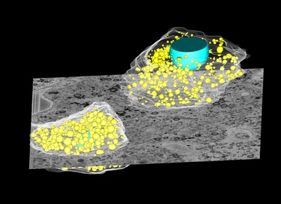
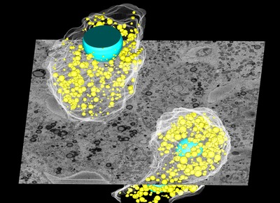
Liver cells under the microscope
Thursday, 12 December, 2013
The purpose of the University of Queensland’s Centre for Microscopy and Microanalysis (CMM), located at the Australian Institute for Bioengineering and Nanotechnology (AIBN), is to promote, support and initiate research and teaching in the applications of microscopy and microanalysis. Appropriately, the centre is now home to Australia’s first in-situ sectioning electron microscope.
CMM Director Professor John Drennan explained that the equipment, worth over $1 million, “was purchased through a university internal competitive scheme, designed to allow researchers to purchase important infrastructure that can be made available to a wide range of users”. It consists of a field emission scanning electron microscope, produced by Zeiss, attached to a Gatan 3View system which includes a diamond microtome knife placed in the sample area.
“The microtome slices and the newly formed surface is imaged by the microscope,” Professor Drennan said. All the scans are collated and a detailed 3D image is built up. The slices can be as small as 60 nm thick, which means the equipment could make 666 slices to cut through an average human hair with a thickness of 40,000 nm.

The equipment has allowed for the publication of a research paper in the journal Current Biology. A group comprising CMM Deputy Director and Institute for Molecular Bioscience lab head Professor Rob Parton, postdoctoral fellow Dr Nicholas Ariotti and University of Barcelona’s Dr Albert Pol used the microscope to view the internal structure of - and interaction between - liver cells.
Professor Parton explained that not all cells are the same. If some cells carry too many lipids (molecules which store fat as energy), they die, while other cells can help the population by taking up large amounts of lipids. The mechanism causing the cells to differ is particularly noticeable in the liver, which Professor Parton says is “a protective social organisation to reduce damage to the liver”. The 3D images generated from the equipment allowed the researchers to see this social organisation, Professor Parton said, and has helped them to understand the importance of cell variation.


The equipment has been used in several other biological and non-biological applications as well, with Professor Drennan saying it is suitable for any materials “that are amenable to be sliced in this way”.
“For example, preliminary results from paint fragments taken from artists’ works have the potential to provide a unique three-dimensional view of the processes that are taking place as the work ages. This information is invaluable for conservators who are always looking to find ways of monitoring and preserving important art works.”
The CMM has always provided students and staff with access to state-of-the-art instrumentation, said Professor Drennan, and the latest acquisition is attracting plenty of attention. Not only are the university’s researchers becoming skilled at using the equipment, but interstate scientists are coming for training as well.
“This new instrument is of interest to a wide range of researchers,” Professor Drennan concluded.
MRI scanner to advance medical breakthroughs at Monash
Siemens Healthineers' MAGNETOM Cima.X 3T is claimed to be Victoria's most advanced,...
Virtual pathology streamlines rapid onsite evaluation
Technology from Grundium, a specialist in digital imaging for pathology, has been shown to match...
Cannabis detected in breath from edibles
Researchers say they have made the first measurement of THC in breath from edible cannabis, in a...


