
Asbestos analysis by transmission electron microscopy, SAED and EDX-S

Due to the detrimental health effects of asbestos, it continues to be a prominent concern, highlighting the need for precise and dependable analytical methods to identify and measure its presence. This article investigates the capabilities of transmission electron microscopy (TEM) in comparison to alternative techniques such as polarised light microscopy (PLM), phase contrast microscopy (PCM) and scanning electron microscopy (SEM).
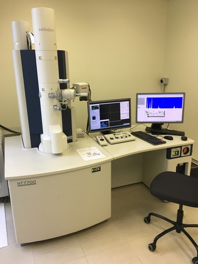
Transmission electron microscope principle
TEM is a highly capable imaging method that allows scientists to examine the internal structure of materials with remarkable resolution. Distinguishing it from alternative microscopy techniques, TEM utilises an electron beam instead of light to visualise the sample. As the electron beam passes through the extremely thin sample, it engages in interactions that provide detailed information about the sample’s composition, crystal structure and morphology.
The analysis commences by meticulously preparing a small section of the asbestos-containing material and placing it onto a copper grid, facilitating its smooth transfer into the vacuum chamber of the microscope. Subsequently, the sample is exposed to a concentrated electron beam, and diverse detectors capture signals that unveil distinct characteristics of the sample’s properties.
Comparing TEM against PLM, PCM and SEM
Within the array of analytical techniques, TEM stands apart due to its unmatched capability to offer comprehensive insights into asbestos fibres. While PLM, PCM and SEM are commonly utilised, they possess specific limitations that TEM successfully overcomes.
PLM
The combination of PLM with dispersion staining relies on the examination of asbestos fibres using polarised light, enabling the identification of fibre type through the analysis of morphological characteristics and crystalline structure.
Nevertheless, PLM may have limitations when dealing with non-commercial types of asbestos or other minerals that possess similar properties, resulting in inconclusive bulk results (referred to as unknown mineral fibres). Additionally, PLM’s resolution limit of 0.2 μm hampers its ability to detect thin and short asbestos fibres that might be present in complex matrices such as vinyl tiles, putty samples, mastics, vermiculite, plasterboard and other non-fibrous materials, and “difficulties may occur in identifying fibres below about 1 μm width” as mentioned in Asbestos: The Analysts’ Guide1.
In both scenarios, TEM’s capacity to detect these concealed asbestos structures guarantees a more thorough and precise evaluation of potential asbestos presence in building materials and natural samples.
PCM
The PCM method, employed for measuring airborne asbestos fibres, lacks the ability to distinguish between asbestos and non-asbestos fibres. As a result, it counts all fibre types in the sample, regardless of whether they are asbestos or not, leading to an overestimation of asbestos fibre concentrations.
Within the Australian regulatory framework, the NOHSC:30032 method utilises PCM to obtain counting results expressed as the number of fibres per millilitre of air. Subsequently, TEM analysis can be performed following the NIOSH 74023 method to identify the ratio in percentage of asbestos fibres on the same filter. This ratio is then applied to the PCM counts to calculate the actual concentration of asbestos fibres present in the air samples.
Nevertheless, PCM is limited by its reliance on light as a source, which poses constraints on its ability to observe objects smaller than 0.2 μm in diameter. Consequently, due to this resolution limit, the combination of results following the NIOSH 7402 method requires TEM to limit its observation exclusively on fibres >0.2 μm in width.
It is also important to note, as stated in ISO 10312 Ambient air — Determination of asbestos fibres — Direct transfer transmission electron microscopy method, “Most fibres in ambient atmospheres are not asbestos and therefore, there is a requirement for fibres to be identified. Many airborne asbestos fibres in ambient atmospheres have diameters below the resolution limit of the optical microscope... [Transmission electron microscopy] has adequate resolution to allow detection of small fibres and is currently the only technique capable of unequivocal identification of the majority of individual fibres of asbestos.”4
Through the utilisation of TEM, it becomes possible to attain a more precise and dependable estimation of asbestos concentration, thereby enhancing risk assessment and ensuring regulatory compliance.

SEM
Conversely, SEM offers high-resolution imaging of asbestos fibres and valuable surface information, in addition to chemical composition input when combined with energy dispersive X-ray spectroscopy (EDX-S), making it an excellent technique to discriminate cleavage fragments from asbestiform fibres of same composition. However, its resolution is limited by its image contrast5,6 capability that may prevent it from visualising thin asbestos fibres. Furthermore, unless equipped with EBSD (electron backscatter diffraction), SEM cannot definitively identify and characterise the internal crystalline structure of the observed fibres, which can lead to result misinterpretation, as demonstrated in cases of talc contaminated with anthophyllite. In such instances, both minerals exhibit similar morphology and chemical composition, making differentiation difficult without examining their internal crystalline structure patterns.

Three criteria for asbestos identification using TEM
TEM relies on three criteria to definitively identify asbestos fibres in both bulk and air samples.
Morphology
The first criterion employed by TEM involves examining the morphology of the observed fibre at high magnification to assess its asbestiform habit. This allows for differentiation between asbestos and non-asbestos fibres based on their characteristic features.

Crystalline structure
The second criterion employed by TEM involves analysing the crystalline structure using selected area electron diffraction (SAED). This technique enables the assessment of fibre anisotropy. Asbestos fibres display a unique pattern of parallel rows with comet tail-like structures, which aids in their classification.

Chemical composition
Lastly, the chemical composition is examined using EDX-S. EDX-S analysis provides crucial information for identifying the precise type of asbestos among the six recognised forms.

Conclusion
The crucial role of TEM in asbestos analysis lies in its ability to provide unique insights into the identification and characterisation of asbestos fibres.
The combination of high-magnification morphology observation (exceeding 10,000x), SAED analysis for anisotropy determination, and EDX-S for chemical composition identification is essential for distinguishing numerous mineral fibres that resemble asbestos but are not hazardous.
TEM’s high resolution and capability to detect thin and short asbestos fibres make it an indispensable tool for accurately assessing building materials, natural samples and air monitoring filters. This capability has contributed to the establishment of efficient standards in sampling, preparing and analysing asbestos by agencies like ISO, NIOSH (USA) and AFNOR (France) since the early 1990s.
In conclusion, the detailed examination of asbestos fibres by TEM establishes it as an indispensable tool for scientists and analysts, enhancing our ability to safeguard public health, provide accurate asbestos-related data, and improve risk evaluation and compliance.
1. https://www.hse.gov.uk/pubns/priced/hsg248.pdf
2. https://www.safeworkaustralia.gov.au/sites/default/files/2021-11/guidancenote_membranefiltermethodforestimatingairborneasbestosfibres_2ndedition_nohsc3003-2005_pdf.pdf
3. https://www.cdc.gov/niosh/docs/2003-154/pdfs/7402.pdf
4. https://www.iso.org/obp/ui/#iso:std:iso:10312:ed-2:v1:en
5. https://echa.europa.eu/documents/10162/4605fc92-18a2-ae48-f977-4dffdecfec11, p29
6. https://www.atsdr.cdc.gov/toxprofiles/tp61.pdf, p188
From assistance to agency in biopharma R&D: benefits of next-gen AI-native lab notebooks
The next generation of AI-native lab notebooks has the potential to turn AI into an active...
Images reveal "incredible" detail of industrial micro-CT scanner
Images of intricate biological structures and everyday items have revealed the capabilities of a...
MRI scanner to advance medical breakthroughs at Monash
Siemens Healthineers' MAGNETOM Cima.X 3T is claimed to be Victoria's most advanced,...


