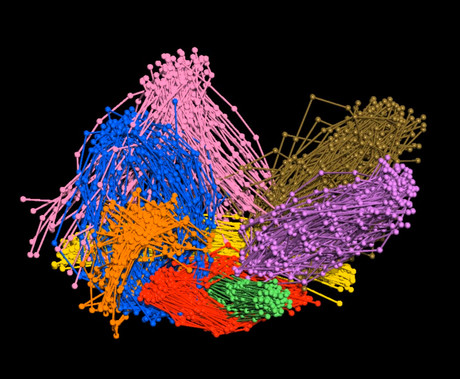
Better in 3D: nanomachines observed in living cells

Through a combination of genetic engineering, super-resolution microscopy and biocomputation, European researchers have been able to observe protein nanomachines in living cells in 3D.
Also known as protein complexes, protein nanomachines are the structures responsible for performing cell functions. Currently, biologists who study the function of these complexes isolate them in test tubes, divorced from the cell, and then apply in vitro techniques that allow them to observe their structure up to the atomic level. They can also analyse these complexes within the living cell, but that provides little structural information.
“The in vitro techniques available are excellent and allow us to make observations at the atomic level, but the information provided is limited,” said Oriol Gallego, who coordinated the study group. “We will not know how an engine works if we dissemble it and only look at the individual parts. We need to see the engine assembled in the car and running.”
Seeking a solution, Gallego and his colleagues at IRB Barcelona, in collaboration with the University of Geneva and the Centro Andaluz de Biología del Desarrollo, developed a strategy that brings together methods from super-resolution microscopy, cell engineering and computational modelling. Their work was published in the journal Cell.
The scientists began by genetically modifying cells in order to build artificial supports inside onto which they could anchor protein complexes. These supports would allow the researchers to regulate the angle from which the immobilised nanomachinery was viewed.
Then, in order to determine the 3D structure of the protein complex, they used super-resolution techniques to measure the distances between different components and then integrate them in a process similar to that used by GPS.
The method enabled the researchers to observe protein complexes with a precision of 5 nm — a resolution four times better than that offered by super-resolution, according to Gallego. By directly observing the structure of the protein machinery in living cells while it is executing its function, the scientists have enabled cell biology studies that were previously unfeasible.

Gallego has already used the method to study exocytosis, a mechanism that the cell uses to communicate with the cell exterior. He and his colleagues have now revealed the entire structure of a key nanomachine in exocytosis that was until now an enigma.
“We now know how this machinery, which is formed by eight proteins, works and what each protein is important for,” said Gallego. “This knowledge will help us to better understand the involvement of exocytosis in cancer and metastasis — processes in which this nanomachinery is altered.”
Furthermore, an understanding of how nanomachines carry out their cell functions has biomedical implications, since alterations in their inner workings can lead to the development of diseases. The new strategy could therefore make it possible to see how viruses and bacteria use protein nanomachines during infection, as well as better understand the defects in complexes that lead to diseases in the first place.
Gallego said the ability to see 5 nm protein complexes is “a great achievement”, though he added that there is “still a long way to go to be able to observe the inside of the cell at the atomic scale that in vitro techniques would allow”.
“I think that the future lies in integrating various methods and combining the power of each one,” he said.
Revealed: the complex composition of Sydney's beach blobs
Scientists have made significant progress in understanding the composition of the mysterious...
Sensitive gas measurement with a new spectroscopy technique
'Free-form dual-comb spectroscopy' offers a faster, more flexible and more sensitive way...
The chemistry of Sydney's 'tar balls' explained
The arrival of hundreds of tar balls — dark, spherical, sticky blobs formed from weathered...


