Chemical imaging for cancerous tissue
Scientists have outlined a new method for analysing biological samples, such as searching for cancer, based on their chemical make-up. The process would transform medical examinations, with tests currently involving interpretation by a histology specialist and a waiting period of weeks to obtain a full result.
Mass spectrometry imaging (MSI) reveals how hundreds or thousands of chemical components are distributed in a tissue sample. Until recently, scientists were yet to devise a method to apply this technique to any type of tissue. Now researchers at Imperial College London have outlined a recipe for processing MSI data and building a database of tissue types, which they have published in Proceedings of the National Academy of Sciences.
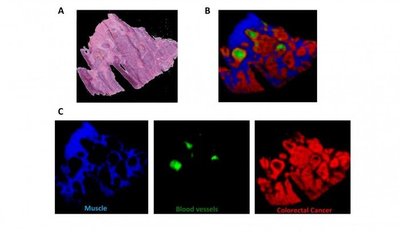
In MSI, a beam moves across the surface of a sample, producing a pixelated image. Each pixel contains data on thousands of chemicals present in that part of the sample. By analysing many samples and comparing them to the results of traditional histological analysis, a computer can learn to identify different types of tissue.
A single test taking a few hours can provide much more detailed information than standard histological tests, eg, showing if a tissue is cancerous and the type and subtype of cancer, which can be important for choosing the best treatment. Corresponding author Dr Kirill Veselkov said MSI is “an extremely promising technology, but the analysis required to provide information that doctors or scientists can interpret easily is very complex”.
The researchers’ solution was an integrated bioinformatics platform, which “allows intuitive histology-directed interrogation of MSI datasets”, they said in the study. They explained, “Unique lipid patterns were observed using this approach according to tissue type, and a tissue recognition system using multivariate molecular ion patterns allowed highly accurate (>98%) identification of pixels according to morphology (cancer, healthy mucosa, smooth muscle and microvasculature).”

Dr Zoltan Takats claims the method is “independent of the user - it’s based on numerical data, rather than a specialist’s eyes - and it can tell you much more in one test than histology can show in many tests”. Professor Jeremy Nicholson added, “Multivariate chemical imaging that can sense abnormal tissue chemistry in one clean sweep offers a transformative opportunity in terms of diagnostic range, speed and cost, which is likely to impact on future pathology services and to improve patient safety.”
The technology will also be useful in drug development. To study where a new drug is absorbed in the body, pharmaceutical scientists attach a radioactive label to the drug molecule, then look at where the radiation can be detected in a laboratory animal. If the label is detached when the drug is processed in the body, it is impossible to determine how and where the drug has been metabolised. MSI would allow researchers to look for the drug and any metabolic products in the body, without using radioactive labels.
“[This] strategy permits the analysis of multivariate molecular signatures with direct correlation to morphological regions of interest, which can offer new insights into how different tumour microenvironmental populations interact with one another and generate novel region-of-interest specific biomarkers and therapeutic targets,” the researchers concluded.
A non-destructive way to locate microplastics in body tissue
Currently available analytical methods either destroy tissue in the body or do not allow...
Rapid imaging method shows how medicine moves beneath the skin
Researchers have developed a rapid imaging technique that allows them to visualise, within...
Fluorescent molecules glow in water, enhancing cell imaging
Researchers have developed a new family of fluorescent molecules that glow in a surprising way,...


