Protein programs killer T cells to migrate to tumour sites

US scientists have discovered how killer T cells ‘learn’ to migrate to sites of infections or tumours, where they amass and attack the diseased cells. Apparently, the answer lies in a protein called Runx3.
During infection or tumour growth, white blood cells called CD8+ T cells rapidly multiply within the spleen and lymph nodes and acquire the ability to kill diseased cells. Some of these killer T cells then migrate where required to vanquish the germs or cancers, amassing within specific tissues like the skin, gut and lung, or solid tumours — and scientists want to know why.
Now, researchers from The Scripps Research Institute and the University of California, San Diego have found that the Runx3 protein programs killer T cells to establish residence in tumours and infection sites. Published in the journal Nature, their work will be useful for developing cancer-fighting immunotherapy strategies.
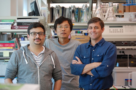
“Runx3 works on chromosomes inside killer T cells to program genes in a way that enables the T cells to accumulate in a solid tumour,” said Matthew Pipkin, an associate professor at The Scripps Research Institute and co-author of the study.
As explained by Pipkin, there are two main strategies in cancer immunotherapy that employ killer T cells. Checkpoint inhibitor blockade unleashes killer T cells, prompting them to accumulate in tumours more aggressively. Adoptive cell transfer, meanwhile, involves reinfusing a patient’s own immune cells after they have been engineered in the lab to recognise and destroy the patient’s specific cancer.
Adoptive cell transfer has worked well in some blood cancers associated with the lymphoid system, according to Pipkin — but there appears to be less efficient activity of T cells in solid tumours. As noted by Pipkin, “The gene programs and signals for how the T cells take up residence in tissues outside of the general circulation was not really well understood.”
To discover factors that control T cell residency beyond the lymphoid system, Pipkin’s team worked with the laboratory of UC San Diego’s Ananda Goldrath, who compared the gene expression of CD8+ T cells found in non-lymphoid tissue to those found in the general circulation. They then employed an RNA interference screening strategy which can test the actual function of thousands of potential factors simultaneously, previously developed in collaboration with the La Jolla Institute for Allergy and Immunology.
“We found a distinct pattern,” Pipkin said. “The screens showed that Runx3 is one at the top of a list of regulators essential for T cells to reside in nonlymphoid tissues.” Moreover, Runx3 was able to engage a specific gene program that is found in natural tissue-resident and tumour-infiltrating CD8+ T cells.
The group further assessed whether Runx3 had a role in directing white blood cells that attack solid tumours in mouse melanoma models. They found that adoptive cell transfer of cancer-specific killer T cells that overexpressed Runx3 delayed tumour growth and prolonged survival, while mouse models treated with those lacking Runx3 fared much worse than normal.
“If we enhance Runx3 activity in the cells, the tumours are significantly smaller and there is greater survival compared to the control group,” Pipkin said.
Knowing that modulating Runx3 activity in T cells influences their ability to reside in solid tumours opens new opportunities for improving cancer immunotherapy, according to Pipkin. “The upshot is we could probably use Runx3 to reprogram adoptively transferred cells to help drive them to amass in solid tumours,” he said.
Neurosensing/neurostimulation implants session to be held on Monday
On Monday, a session at UNSW Sydney will include people who are benefiting from bioelectronics...
argenx and Monash University partner against autoimmune diseases
To advance a pioneering molecule for autoimmune diseases, global immunology company argenx has...
Archer completes potassium sensing alpha prototype
Quantum technology company Archer Materials Limited has developed an early Biochip prototype...


