Images of insulin in action could lead to better diabetes treatments
An Australian-led research team has obtained the world’s first 3D pictures of insulin in the process of binding to cell surfaces so that the cells can take up sugar from the blood. The images solve the 20-year mystery of how insulin binds to the insulin receptor and will enable the development of improved forms of insulin for treating type 1 and 2 diabetes.
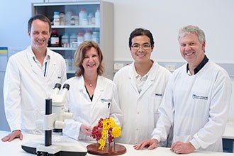
A research team led by Associate Professor Mike Lawrence, Dr Colin Ward and Dr John Menting from the Walter and Eliza Hall Institute of Medical Research in Melbourne used X-ray diffraction at the Australian Synchrotron to obtain highly detailed, three-dimensional images of insulin and the insulin receptor. Their work has been published in the prestigious scientific journal Nature.

Associate Professor Lawrence said the team was excited to reveal for the first time a three-dimensional view of insulin bound to its receptor.
“The insulin receptor is a large protein on the surface of cells to which the hormone insulin binds,” he said.
“Understanding how insulin interacts with the insulin receptor is fundamental to the development of novel insulins for the treatment of diabetes.
“We can now exploit this knowledge to design new insulin medications with improved properties, which is very exciting.”
The synchrotron X-ray images show that both insulin and the insulin receptor change their shape in order to bind with each other, in a manner which Associate Professor Lawrence describes as “unusual”.
“Both insulin and its receptor undergo rearrangement as they interact - a piece of insulin folds out and key pieces within the receptor move to engage the insulin hormone,” he said. “You might call it a ‘molecular handshake’.”
Associate Professor Lawrence said the work would not have been possible without access to the Australian Synchrotron’s specialised MX2 microfocus beamline and the synchrotron MX team led by Dr Tom Caradoc-Davies, who kept the beamline operating at world-class standard.
“If we did not have this fantastic facility in Australia and their staff available to help us, we would simply not have been able to complete this project,” Associate Professor Lawrence said.
The microfocus beamline has a highly focused X-ray beam that allows scientists to collect useful diffraction data from very small crystals such as those used in the insulin work, which were comparable to the width of a human hair. The beam is thousands of times brighter than laboratory X-ray sources, enabling experiments to be completed in minutes rather than weeks.
“It’s difficult to grow crystals of insulin docking with its receptor, but frequent synchrotron visits meant we could optimise the crystal preparation methods and the experimental settings rather than guessing what might work,” Associate Professor Lawrence said.
Further project collaborators included researchers from Case Western Reserve University, the University of Chicago, the University of York and the Institute of Organic Chemistry and Biochemistry in Prague.
“Collaborations in this field are essential,” Associate Professor Lawrence said. “No one laboratory has all the resources, expertise and experience to take on a project as difficult as this one.”
With approximately one million Australians currently living with diabetes and around 100,000 new diagnoses each year, it is vital that new methods of treatment are sought.
“This discovery could conceivably lead to new types of insulin that could be given in ways other than injection, or an insulin that has improved properties or longer activity so that it doesn’t need to be taken as often,” Associated Professor Lawrence said.
“It may also have ramifications for diabetes treatment in developing nations, by creating insulin that is more stable and less likely to degrade when not kept cold, an angle being pursued by our collaborators.”
The project was supported by the National Health and Medical Research Council of Australia and the Victorian Government.
For more information, watch the video below.
Why are young plants more vulnerable to disease?
Fighting disease at a young age often comes at a steep cost to plants' growth and future...
Liquid catalyst could transform chemical manufacturing
A major breakthrough in liquid catalysis is transforming how essential products are made, making...
How light helps plants survive in harsh environments
Researchers from National Taiwan University have uncovered how light stabilises a key...


