
Extend the life and performance of your microscope — without breaking the bank
By Craig Rappaport, Senior Sales Specialist, Life Science Microscopy*
Tuesday, 19 April, 2022

The bounds of research microscopy are rapidly expanding while individual lab budgets are shrinking.
Does your lab wish to pursue new microscopy applications but you have a limited budget and an older system? Is an expensive new instrument necessary to facilitate the research or can an existing microscope be reinvigorated or upgraded to meet near-term needs?
This article presents ways to squeeze additional performance out of an older microscope or to even repurpose the system for new application requirements. If these ideas spark interest, consult a local microscopy expert to find out how much life is left in that old workhorse. As one might expect, making sure the instrument is in good condition and has appropriate optics are two of the most important considerations. Other components, such as filters and digital cameras, can significantly improve image data acquisition.
1. Preventive maintenance
As with any major piece of equipment, the first thing one should do before investing in modernisation is to make sure the ‘bones’ are in good shape. High-quality light microscopes can go for several years without appearing to need major service. What often goes unattended are simple things like basic cleaning and lubrication. Oil immersion objectives, if not properly cleaned, can produce ‘soft’ images. Likewise, non-immersion (‘dry’) optics may inadvertently be dragged through oil left on a slide, resulting in blurry images. Understanding how to safely clean your optics is as important as knowing how to change your microscope’s bulbs. Many manufacturers offer resource guides in PDF or online format. Alternatively, some facilities have microscopy cores or specialists to provide instruction.
Focusing binocular tubes and oculars can freeze if not lubricated, stages become ‘grindy’, focus mechanisms become stiff and iris diaphragms bind up. These different parts of the microscope can require different types of lubricants. A qualified microscope service technician can apply fresh oils and can often repair or replace broken components onsite. In cases of severe disrepair, many service companies can perform a full microscope overhaul in their shop. Many local service companies work on multiple brands. If you’re not sure who to contact, submit a support request through your vendor’s service/support website. Once the microscope is operating smoothly, one can begin to consider where to invest next.
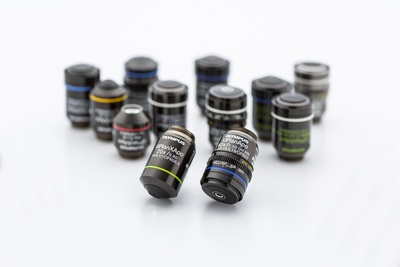
2. Upgrade your objectives
The objectives are the heart of your microscope system. Selecting application-specific optics is fundamental for excellent image data. Even if the microscope was purchased as recently as five years ago, there is a strong possibility the manufacturer has made technological improvements returning higher resolution, better chromatic aberration, flatter images across the field of view (both in terms of focus and fluorescence intensity), longer working distances, etc.
In addition, silicone oil objectives with a refractive index (RI) matched to one’s biological samples maintain viscosity and RI at temperature, produce more accurate volume rendering and offer longer working distances than standard immersion oil objectives. These can be highly beneficial for time-series live cell imaging.
Depending on the manufacturer, newly designed objectives may be backward-compatible to instruments even 20+ years old. To identify whether a system is infinity corrected, look for the infinity (∞) symbol on the objective barrel. If uncertain, consult a microscope specialist.
Some manufacturers may still offer discontinued older inventory not shown on their website, so it is worth asking the local sales representative. When a manufacturer’s current optics are not compatible with an older instrument an online search may help identify alternative sources. This can be particularly helpful for discontinued 160 mm microscopes (these will typically have ‘160’ printed on the objective barrel to denote a 160 mm tube length). Of course, one must be careful to find out if used equipment carries a warranty, whether there is a return policy or if the items are sold ‘as is’.
3. Replace old fluorescence filters
Older fluorescence filters (soft-coated or laminated filters) diminish in efficiency within a few hundred hours of exposure to high-intensity light. Furthermore, over time they would burn or delaminate, becoming even less efficient and potentially shifting their spectral characteristics.
Current filter manufacturing technology has virtually eliminated wear-out and burnout problems. If you know how, you can pull your fluorescence filter cube or slider from your microscope and, by tilting it at an angle, inspect your excitation and emission filters for signs of wear. Most commonly, you will notice mottling on the filter and/or a ‘hole’ where excitation light has burned out the centre area of the filter. If not sure, a microscope technician or sales specialist can examine the filters and make recommendations based on their condition. If you are planning on purchasing a new digital camera, ask the salesperson to check your filters first.

4. And about that old digital camera…
By now most IT departments have mandated upgrading Windows PCs to Windows 10, or even 11. Depending on the age of one’s system, an older digital camera could be obsolete due to lack of support in the current version of Windows. If it’s been a while since a camera was purchased, one will find that a lot has changed. Modern sensors offer higher megapixels, faster frame rates and greater sensitivity than ever before. But is a colour camera required, or monochrome, or both?
If the applications involve chromogenic stains such as hematoxylin and eosin (H&E), a colour camera is required. If the work is in fluorescence imaging, a sensitive colour camera may suffice, so long as the emission wavelengths don’t run into the infrared (IR) (such as with Cy5 or Cy7). This is because colour cameras are filtered for visible wavelengths and thus have much reduced sensitivity beyond 700 nm.
For multichannel fluorescence imaging, especially with live cells and over time, a sensitive monochrome camera is the best choice. These have broad spectral sensitivity and thus can produce better results with shorter exposure times to limit phototoxicity, bleaching and other negative effects of fluorescence illumination.
For those with uncompromising requirements for both colour and monochrome cameras, it may be possible to purchase a dual camera adapter for your microscope. If not, there’s always the less-than-optimal choice of switching them out as needed. In this event, great care must be taken to avoid getting dust on the camera sensor area.
As outlined above, updating or repurposing one’s microscope does not necessarily mean a huge expense. And once the basics have been dealt with, one might consider additional upgrades such as a motorised stage, focus drive, environmental enclosure for temp/CO2/humidity control and modern software for running all your devices. So long as ‘the bones’ are still solid, there likely is plenty of capability left in that ageing but robust microscope.

Please follow us and share on Twitter and Facebook. You can also subscribe for FREE to our weekly newsletters and bimonthly magazine.
Benchtop NMR used to assess heart disease risk
The breakthrough will enable more accessible, high-throughput cardiovascular disease (CVD) risk...
Activated gold helps visualise drug movement in the body
Gold nanoparticles are promising drug carriers for cancer therapy and targeted drug delivery, but...
Plasma-modified graphene enhances gas sensors
Researchers have improved gas-sensing technology by treating graphene sheets with plasma under...


