
MRI enables early detection of Alzheimer's in mouse brains

Early identification of Alzheimer’s disease gives an opportunity to halt or slow down its progression into a devastating illness that affects the daily life of patients — but diagnosis of the disease is not easy due to its overlapping signs with normal ageing.
Now, an international research team led by City University of Hong Kong (CityU) has developed a non-invasive molecular imaging approach based on magnetic resonance imaging (MRI) to dynamically measure glucose level changes in the brain’s lymphatic system. Published in the journal Science Advances, their discovery may help in identifying Alzheimer’s disease at early stages so that treatment can begin as soon as possible.
“The tricky part of identifying Alzheimer’s disease is that early abnormalities, such as the emergence of protein plaques, are similar to normal ageing in the human brain,” said CityU’s Dr Kannie Chan Wai-yan. “Patients diagnosed with symptoms, in which deposition of plaques are found in their brain hampering the cognitive function, are most likely already in the middle or late stage of the disease. Actually, pathologies in the brain happened 15 or 20 years before the symptoms appear.”
Recent findings have shown that abnormal clearance of waste in the glymphatic system — the lymphatic system in the brain — is one of the hallmarks of early Alzheimer’s disease. The glymphatic system is a drainage-like system for cerebrospinal fluid to flow through the brain tissue called brain parenchyma, thus facilitating efficient clearance of solutes such as glucose and protein waste from the brain.
Currently, glucose uptake and metabolism can be assessed by imaging with positron emission tomography (PET) in hospitals. However, PET scans with radioactive tracers are expensive, and their invasive nature has hindered general clinical application. Moreover, patients cannot be scanned too frequently with radioactive tracers.
Seeking a more appropriate alternative, Dr Chan’s team developed a new imaging approach based on chemical exchange saturation transfer MRI (CEST MRI) to assess glucose uptake and clearance in the glymphatic system of the mice’s brains non-invasively. “CEST MRI has been used in the diagnosis of brain tumours,” Dr Chan revealed, “[but] this is the first time that it is used in assessing the function of glymphatic system.”
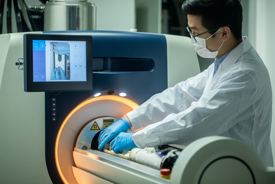
The team carried out the experiments using a 3T MRI animal scanner at CityU, said to be the only of its kind in Hong Kong. They injected glucose into genetically modified mice with Alzheimer’s disease and healthy mice aged six months and 16 months. The dynamic response of glucose — both in the cerebrospinal fluid and brain parenchyma — was then measured using the CEST MRI.
According to the MRI results, the Alzheimer’s disease mice showed significantly slower cerebrospinal fluid clearance rates than the age-matched healthy mice, which is consistent with previous neuropathological findings. “Clearance rates are reduced because of abnormalities in the brain’s drainage system,” Dr Chan explained.
Moreover, significantly higher glucose uptake was detected in brain parenchyma of the six-month-old mice with Alzheimer’s disease compared to the healthy mice of the same age. For the 16-month-old mice with Alzheimer’s disease, significantly lower glucose uptake was found in both brain parenchyma and cerebrospinal fluid compared to the age-matched healthy mice. These results echo previous research findings using other methodologies, and also serve as hallmarks to differentiate Alzheimer’s disease from normal ageing.

The imaging of glucose uptake and clearance in the cerebrospinal fluid and brain parenchyma thus enables the assessment of the brain glymphatic system. Importantly, abnormalities are detected at the early stage of Alzheimer’s disease when little neuropathology develops in the brain. The team believes this non-invasive assessment of the glymphatic system can serve as an imaging biomarker to reveal the early pathology in Alzheimer’s disease.
“By using glucose as a ‘tracer’, our imaging method can sensitively detect the distinctive changes of glymphatic system function at the molecular level at an early stage of the disease, helping us to differentiate it from normal ageing,” Dr Chan said. “Besides, glucose is natural, biodegradable and commonly used in hospitals, such as the glucose tolerance test. Using it as a contrast agent for MRI is non-invasive and safe.”
She added that the new imaging method is compatible with the MRI machines commonly used in clinics or hospitals, which should mean low set-up cost and easy transfer into clinical application.
Having successfully identified changes in the brains of mice using a 3T MRI scanner, Dr Chan said she expects changes in the human brain to be more detectable since human brains are much larger. She anticipates that clinical trials could be conducted within three years.
Please follow us and share on Twitter and Facebook. You can also subscribe for FREE to our weekly newsletters and bimonthly magazine.
Benchtop NMR used to assess heart disease risk
The breakthrough will enable more accessible, high-throughput cardiovascular disease (CVD) risk...
Activated gold helps visualise drug movement in the body
Gold nanoparticles are promising drug carriers for cancer therapy and targeted drug delivery, but...
Plasma-modified graphene enhances gas sensors
Researchers have improved gas-sensing technology by treating graphene sheets with plasma under...


