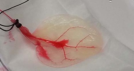
Human heart tissue grown in spinach leaves

US scientists may have solved a major tissue bioengineering problem, using the vascular system of spinach leaves to support human heart cells. Published in the journal Biomaterials, their study opens the door to eventually growing layers of healthy heart muscle to treat heart attack patients.
While researchers have previously succeeded in regenerating small samples of human tissue, scaling up this tissue to full size brings with it certain complications. Current bioengineering techniques, including 3D printing, cannot fabricate the branching network of blood vessels that are required to deliver the oxygen, nutrients and essential molecules required for proper tissue growth.
“The major limiting factor for tissue engineering a graft and getting it to be into the clinic is the lack of a vascular network,” explained Joshua Gershlak, first author on the new paper.
“Without that microvasculature, you lose that oxygen transport. And so as you build a bigger and bigger graft, for say like a heart attack on a human, you’re going to need something fairly large. So without that vascular network, you get a lot of tissue death.”
To solve this problem, Gershlak and a multidisciplinary research team at Worcester Polytechnic Institute (WPI), the University of Wisconsin-Madison and Arkansas State University turned to plants.
“Plants and animals exploit fundamentally different approaches to transporting fluids, chemicals and macromolecules, yet there are surprising similarities in their vascular network structures,” the study authors wrote. “The development of decellularized plants for scaffolding opens up the potential for a new branch of science that investigates the mimicry between plant and animal.”
WPI’s Glenn Gaudette, corresponding author on the paper, explained that the researchers took spinach leaves and perfused them with a detergent that removed all their cellular material. This process was developed by Gershlak, a graduate student in Gaudette’s lab.
“I had done decellularisation work on human hearts before, and when I looked at the spinach leaf its stem reminded me of an aorta,” said Gershlak. “So I thought, let’s perfuse right through the stem. We weren’t sure it would work, but it turned out to be pretty easy and replicable.”
Once the plant cells were washed away, what remained was a framework made primarily of cellulose — the structure that normally keeps those cells in place.
“Cellulose is biocompatible [and] has been used in a wide variety of regenerative medicine applications, such as cartilage tissue engineering, bone tissue engineering, and wound healing,” the authors wrote. Seeding human cells onto this structure was therefore not going to be a problem.
“The idea here is that we have this very thin, flat piece of tissue that already has a vascular network in there, and so we should be able to potentially stack up multiple leaves and create a piece of cardiac tissue,” said Gershlak.
The scientists flowed fluids and microbeads similar in size to human blood cells through the spinach vasculature, then seeded the spinach veins with human mesenchymal stem cells and pluripotent stem cell-derived cardiomyocytes. Gershlak said these cells both attached to and contracted on the spinach leaf, looking and acting “like normal cardiac cells”.
“We can in theory sew those veins into the native arteries in the heart and therefore produce a contractile muscle that can replace the infarct or the dead tissue in the heart [of heart attack victims],” added Gaudette.

The researchers noted that other decellularised plants could provide the framework for a wide range of tissue engineering technologies, with their cell removal technique also found to succeed in parsley, Artemesia annua (sweet wormwood) and peanut hairy roots.
“The spinach leaf might be better suited for a highly vascularized tissue, like cardiac tissue, whereas the cylindrical hollow structure of the stem of Impatiens capensis (jewelweed) might better suit an arterial graft,” the authors wrote. “Conversely, the vascular columns of wood might be useful in bone engineering due to their relative strength and geometries.”
According to Gaudette, the researchers are now seeking to optimise the decellularisation process and to further characterise how various human cell types grow while they are attached to, and are potentially nourished by, plant-based scaffolds. They will also explore the option of engineering a secondary vascular network for the outflow of blood and fluids from human tissue.
Blood test for chronic fatigue syndrome developed
The test addresses the need for a quick and reliable diagnostic for a complex,...
Droplet microfluidics for single-cell analysis
Discover how droplet microfluidics is revolutionising single-cell analysis and selection in...
PCR alternative offers diagnostic testing in a handheld device
Researchers have developed a diagnostic platform that uses similar techniques to PCR, but within...


