Nanoparticles, peptides and paper to detect cancer
Engineers at Massachusetts Institute of Technology (MIT) have developed a cheap and simple diagnostic, much like a pregnancy test, which detects cancer in urine samples. The team described their technology in Proceedings of the National Academy of Sciences.
“With non-communicable diseases (NCDs) now constituting the majority of global mortality, there is a growing need for low-cost, non-invasive methods to diagnose and treat this class of diseases, especially in resource-limited settings,” the researchers said. They added that NCDs are “challenging to diagnose because biomarkers that can accurately detect them in patients are lacking”.
In 2012, Professor Sangeeta Bhatia and her MIT colleagues introduced the concept of synthetic biomarker technology to amplify signals from tumour proteins that would be hard to detect on their own. These proteins - proteases known as matrix metalloproteinases (MMPs) - help cancer cells escape their original locations by cutting through proteins of the extracellular matrix.
The researchers explained that their synthetic biomarkers are “composed of nanoparticles conjugated to ligand-encoded reporters via protease-sensitive peptide substrates”. After being injected into the patient, the particles target tumour sites where the MMPs “cleave the peptide substrates and release reporters that are cleared into urine”.

Professor Bhatia noted that when the researchers originally invented the synthetic biomarker, they used a mass spectrometer to conduct the analysis. “For the developing world,” she said, “we thought it would be exciting to adapt it instead to a paper test that could be performed on unprocessed samples in a rural setting, without the need for any specialised equipment.”
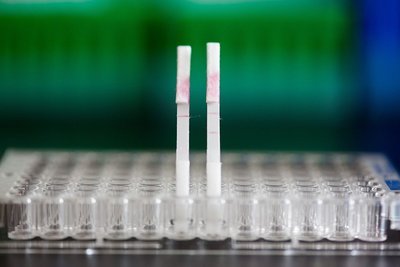
The researchers adapted the particles so they could be analysed on paper, using an approach known as a lateral flow assay (LFA) - the same technology used in pregnancy tests. They first coated nitrocellulose paper with antibodies that can capture the peptides. Once captured, the peptides flow along the strip and are exposed to several invisible test lines made of other antibodies specific to different tags attached to the peptides. If one of these lines becomes visible, it means the target peptide is present in the sample.
“This platform does not require expensive instruments, invasive procedures or trained medical personnel, and may allow low-cost diagnosis of diseases at the point of care in resource-limited settings,” the researchers said. Furthermore, the test takes only minutes and can be modified to detect multiple types of peptides released by different types or stages of disease.
The technology has already been tested in mouse models to detect colorectal cancer and thrombosis. The team is now hoping to identify signatures of MMPs that could be exploited as biomarkers for other types of cancer, as well as for tumours that have metastasised. They are also working on a nanoparticle formulation that could be implanted under the skin, rather than injected, for long-term monitoring.
The research team recently won a grant from MIT’s Deshpande Center for Technological Innovation to develop a business plan for a start-up that could work on commercialising the technology and performing clinical trials. Professor Bhatia says the technology could be applied in high-risk populations, developing nations and even in developed nations as a simple and inexpensive alternative to imaging.
Droplet microfluidics for single-cell analysis
Discover how droplet microfluidics is revolutionising single-cell analysis and selection in...
PCR alternative offers diagnostic testing in a handheld device
Researchers have developed a diagnostic platform that uses similar techniques to PCR, but within...
Urine test enables non-invasive bladder cancer detection
Researchers have developed a streamlined and simplified DNA-based urine test to improve early...


