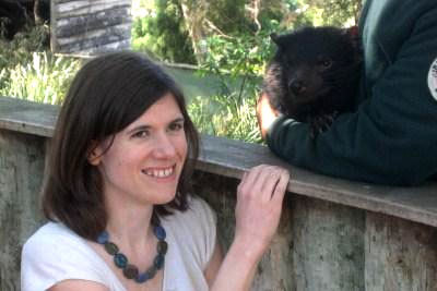
Feature: How next generation sequencing could save the Tasmanian devil

This feature appeared in the May/June 2010 issue of Australian Life Scientist. To subscribe to the magazine, go here.
The devil facial tumour disease (DFTD) is a rare type of infectious cancer that threatens to rid Australia of one of its most iconic marsupials: the cute (sort of), and feisty (definitely), Tasmanian devil (Sarcophilus harrisii). Placed on the endangered list in 2008, the Tassie devil is in big trouble; at current levels of population decline and disease spread, the devils could go the way of the Tasmanian tiger within two or three decades.
The facial tumour grows very rapidly on the face or inside the mouth of affected devils. Death usually ensues just months after the appearance of initial symptoms, possibly by obstructing the devils’ ability to feed. First observed in 1996, the cancer has spread swiftly through the Tasmanian devil population and there is no treatment for the disease.
DFTD is transmitted by the physical transfer of living cancer cells through biting, which, unfortunately, is a signature part of this carnivorous scavenger’s natural behaviour. This mode of transmission is, by all records, extremely rare, with only one other known tumour spread in a similar way: a sexually transmitted disease of dogs called canine transmissible venereal tumour.
Murchison has pondered extensively on why her beloved devils have been hit by such a rare type of cancer. “The very odd thing is that tumour cells can survive in allogeneic hosts, that is, different individuals. I think there is definitely an element of random chance in it, but probably a larger contribution is the isolation of the devil population,” she says. “The devil population has low levels of genetic diversity, which may have allowed this tumorigenic graft to take hold and travel through the population without being detected by the immune system.”
Characterising cancer
While doing her PhD in genetics at Cold Spring Harbor, in the Unites States, Murchison became interested in DFTD and in understanding the tissue of origin of the disease. Having always had an interest in genome sequencing, she was attracted to the challenge of genetically characterising this relatively new cancer and of Tasmanian devil genomics in general.
“Marsupials are an understudied group of mammals in terms of genomics, and there are basically no tools at all available for this species. Luckily, new sequencing techniques and advances have allowed us to take on this sort of challenge in a way that would not have been possible even two or three years ago.”
---PB---
Murchinson’s interest grew into a body of work that would not only launch her scientific career big time, but also prove extremely valuable in the ongoing fight to learn more about DFTD and thus increase the chance of a cure.
The first step was to look at microRNAs (miRNAs) in tumour samples from devils. These are small, non-coding RNAs that are often highly conserved among species, such that their tissue expression pattern can serve as a tissue marker in some cases. miRNA profiles have previously been used in human cancers to determine the tissue of origin of an unknown primary tumour. In addition, Murchison already had a lot of experience with miRNA sequencing from her PhD work.
The team set about cloning and sequencing all the miRNAs from a range of Tasmanian devil tissues (kidney, heart, spleen, brain, etc.) as well as from the tumour, using state-of-the-art high-throughput DNA sequencing technologies. These sequences were annotated and compared to work out which devil tissue the tumours most resembled. The devil tumour clustered strongly with brain, which accorded with separate work done in the Tasmanian Animal Health labs that identified neural-type protein markers in tumour samples.
With this first hint of a tissue of origin, Murchison and colleagues undertook more in-depth sequence analyses by looking at the transcriptome of the tumour, which would give them a map of gene activity within the tumour cells.
“We sequenced the transcriptomes of tumour and testis from the same individual. We had no real tools at the time for the devil sequencing, thus we chose testis because it expresses a large diversity of genes that would help us annotate the largest number of genes from the tumour sample.”
The two transcriptomes were annotated based on conservation, so gene identities were assigned to the expressed devil tumour sequences in the transcriptome compared to known genes from humans and from opossum, the closest completed marsupial genome. Around 14,000 genes were expressed in both the normal tissue and in the tumour, but their profiles were clearly different.
“We then did a basic subtraction analysis,” says Murchison. “This compared the profiles of genes between the testis and tumour gene libraries and looked for genes that were more highly abundant in a specific and significant way in the tumour relative to the testis.”
Surprising findings
The results that emerged were a bit of a surprise to all involved in the project. The tumour expressed genes that matched strikingly with genes controlling nerve fibre myelination – a job carried out by oligodendrocytes in the central nervous system and by Schwann cells in the periphery.
Murchison did not believe the results at first, but further analysis of the relevant genes and validation of their expression by other methods such as Q-PCR and immunohistochemistry confirmed it.
---PB---
“The myelin-related genes that were abundant in the tumour were very closely related to the myelin genes expressed in Schwann cells, and not to those in oligodendrocytes,” she says. The only conclusion from such compelling evidence was that the DFTD had arisen from a Schwann cell or Schwann cell precursor, and Murchison was finally convinced.
Further immunohistochemistry revealed that a couple of these myelin-related genes are incredibly specific for the tumour, with their expression showing up only in the tumour tissue and in Schwann cells around nearby nerve fibres. This finding became the basis for a diagnostic test using those genes as markers for the tumour.
“This test is more specific than anything else we had, and it is very rapid and simple to do,” says Murchison. It will enable detection of the cancer at an early stage and also allow scientists to distinguish DFTD from other similar cancers that afflict Tasmanian devils. These genetic markers could also help research into the biology of the devil cancer and into more targeted prevention efforts.
The transcriptome analysis also hinted at potential immunosuppressive factors being secreted by the devil tumour, although these findings are yet to be validated physiologically. “I think it is not simply the case that devils are very genetically similar and so cannot recognise the cancer graft as being foreign,” Murchison explains. “I would not be surprised if the tumour is actively preventing or suppressing immune detection by the host. Of course, much more work is needed to even start understanding this aspect of the disease biology.”
This body of work, published earlier this year in Science, was an international team effort. Led by Murchison, it involved labs and people from Canberra, Melbourne, Tasmania and multiple sites in the US, with funding from several major research funding agencies across both countries, as well as Roche, which supplied the 454 sequencing instrumentation.
Murchison did her work at Cold Spring Harbor and in Professor Jenny Graves’ Comparative Genomics Research Group at The Australian National University in Canberra. Many of the samples of course were from Tasmania and she had significant input from bioinformatics experts in Melbourne, led by one of Murchison’s main collaborators, Dr Tony Papenfuss at the Walter and Eliza Hall Institute.
Last year, Murchison moved to the Wellcome Sanger Trust Institute, near Cambridge in the UK, funded by a four year NHMRC Overseas Postdoctoral Biomedical Fellowship and L’Oreal-UNESCO For Women in Science UK and Ireland Fellowship to study devil genomics. She is part of the high-profile Cancer Genome Project, which aims to sequence the genomes of several different cancers and characterise their mutation profiles.
“Being part of the Cancer Genome Project has given me a fantastic opportunity to help the devil,” Murchison says. “DNA sequencing is becoming faster and cheaper, which is fortunate as we are sort of running out of time on this one.”
Murchison graduated from Melbourne University and went on to start her PhD at Cold Spring Harbor in 2002, looking at gene silencing by miRNAs. On hearing from friends about this new disease in Tasmanian devils and how serious a threat it was, she had some tumour samples sent over from Tasmania, just to play with during her PhD. This quickly grew into a full-time project, and grants from a number of sources enabled her to investigate Tasmanian devil genomics.
This feature appeared in the May/June 2010 issue of Australian Life Scientist. To subscribe to the magazine, go here.
AI-designed DNA switches flip genes on and off
The work creates the opportunity to turn the expression of a gene up or down in just one tissue...
Drug delays tumour growth in models of children's liver cancer
A new drug has been shown to delay the growth of tumours and improve survival in hepatoblastoma,...
Ancient DNA rewrites the stories of those preserved at Pompeii
Researchers have used ancient DNA to challenge long-held assumptions about the inhabitants of...


