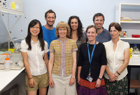
Feature: Stem cell therapy targets Hirschsprung’s disease

This issue appeared in the January/February 2012 edition of Australian Life Scientist. To subscribe, click here.
At the Berlin Congress for Children’s Diseases in 1888, Denmark’s first paediatrician, Harald Hirschsprung, described the deaths of two infants from a rare disorder involving intractable constipation and severe distension of the colon.
The children born with the disorder, which came to bear his name, suffer from a failure of peristalsis, the rhythmic, wave-like sequence of contractions and relaxation that moves the colon’s contents to the rectum for elimination.
More than 50 babies are born in Australia each year with Hirschsprung’s disease, and require surgery to repair a paralysed lower colon. Surgery for the once lethal disorder is life-saving, but the results are not always ideal.
Thus, University of Melbourne neuroscientist Associate Professor Heather Young confesses to having been being “a bit of a sceptic” when Japanese paediatric surgeon Dr Ryo Hotta suggested to her neural stem cell therapy could be a permanent cure for Hirschsprung’s disease.
But when he asked to come and do a PhD project in her laboratory in the Department of Anatomy and Cell Biology, Young was happy to take up his offer. Her team was short of hands, and his idea at least piqued her curiosity.
Like Hirschprung, Young has devoted a large part of her career to the study of the gut, in particular the genesis of the nerve networks that control gut function. Newborns with Hirschsprung’s disease lack the full complement of neurons that enable the lowermost colon to undergo peristalsis. It appears the neuron precursors (neural crest cells) don’t migrate far enough to colonise the colon’s full length during embryonic development.
The journey from the hindbrain to the distal end of the human gut, which continues to elongate ahead of the approaching neural crest cells, takes three weeks, and is probably the longest journey that neural crest cells undertake in their tour of duty innervating the major organs during embryonic development.
Without surgical treatment, Hirschsprungs patients suffer severe constipation that blocks paralysed segment of the lower colon, and distension backs up along the healthy colon. Surgery involves removing the affected segment of the lower colon, drawing down the healthy section, and anastomosing it to the rectum.
But patients tend to suffer continuing gut motility problems involving soiling or incontinence when neurons on either side of the junction fail to make appropriate connections between the colon and the sphincter muscles.
Neurons are essential for motility in all regions of the gut, from oesophagus (top) to anus (bottom). Like Hirschsprung’s patients, diabetic patients commonly suffer from gastroparesis in which there is delayed emptying of the stomach, and aged people affected by Parkinsons disease or achalasia, an oesophageal disorder that affects the swallowing reflex, suffer from motility disorders due to the loss of nerve cells in the gut.
---PB---
Scepticism unfounded
However, Young is looking forward to informing her colleagues at the Australian Neuroscience Society’s annual conference on the Gold Coast in late January that her scepticism about Hotta theory was thoroughly unfounded.According to Young, the variable length of constricted colon in Hirschsprungs patients, which is a consequence of a lack of nerve cells, can be caused by disruption to a variety of signalling pathways that guide neural crest cells down the developing gut during embryogenesis. These signalling pathways control the survival, proliferation, migration and differentiation of neural crest cells.
As well as differentiating into the neurons of the gut, neural crest cells also give rise to the full complement of neurons of the peripheral nervous system, sympathetic and parasympathetic. They also form of much of the connective and skeletal tissues of the head, and the melanocytes that give pigment to the skin.
Mutations in more than a dozen genes, and several uncharacterised genetic loci, have been implicated as risk factors for Hirschsprung’s disease. The major Hirschsprung’s susceptibility gene is RET, which encodes a receptor tyrosine kinase. Mutations in the gene encoding glial cell-derived neurotrophic factor (GDNF), which is the ligand for RET, have also been associated with some cases of Hirschsprung’s disease.
As victims of the colon’s caprices can attest, the organ has a ‘mind’ of its own, which enables it to function independently of the brain.
Young says the digestive tract actually contains more neurons than the spinal cord: around 15-20 subtypes, including sensory neurons that monitor distension of the gut or the presence of nutrients within the lumen, interneurons that project anally or orally and coordinate the directionality of peristalsis, and motor neurons that regulate the contraction and relaxation of the muscle forming the wall of the bowel.
Gut neurons express many of the same neurotransmitters and neurotransmitter receptors that are expressed in the brain, so some brain-active drugs can affect drug motility.
For example, codeine-containing analgesics can have unpleasant side effects on gut motility, while selective serotonin reuptake inhibitors (SSRIs), which are primarily used to treat depression, are also sometimes prescribed to treat irritable bowel syndrome.
---PB---
Mouse model
Hideki Enomoto from the RIKEN Center for Developmental Biology in Kobe, Japan, who is a long-time collaborator of Young’s, engineered mice to express green fluorescent protein in their neural crest cells under the control of the RET gene’s promoter.The green glow reveals the migratory paths of the neural crest cells from the hindbrain down into the undifferentiated mesenchymal tissues of the gut as it elongates during embryogenesis.
These mice have proven crucial for Young’s research. “The gut forms from an inner layer of epithelial cells, while the mesenchymal cells that surround it form multiple layers that differentiate into smooth muscle and connective tissues.
“The undifferentiated mesenchymal tissue secretes factors, including GDNF. In collaboration with Dr Don Newgreen, head of the embryology research group at the Murdoch Children’s Research Institute in Melbourne, we showed that GDNF is chemoattractive to RET-expressing neural crest cells in culture.
“Later, we did an experiment to see if the neural crest cells behave if there is a gradient of GDNF or other chemoattractants, and found that they migrated happily in both directions, so it seems that GDNF promotes cell motility in a non-directional manner.”
In essence, the cells move towards the lower end of the gut because they sense a population deficit beyond the advancing wave front of differentiating neural crest cells. “Cell-to-cell interactions between neural crest cells are also important for their migration down the gut.”
Working with her colleague, Dr Richard Anderson from Anatomy and Cell Biology, who studies the role of cell adhesion molecules (CAMs) in the development of the nervous system, Young’s team used an in vitro technique for growing explants of embryonic gut to examine the role of a CAM called L1.
“When we perturbed the function of L1, less cell-to-cell contact between migrating neural crest cells was observed and migration was retarded,” says Young. “We also identified an inhibitory cue for migration, called semaphorin 3A, which may be the stop signal when the migrating cells reach the end of the gut.
“A sub-population of neural crest cells begin to differentiate into neurons that have neurites that extend in the same direction as the migrating cells. Imaging of the fluorescent cells suggests the neurites provide a substrate that enables the migrating cells just behind the wavefront to migrate fastest. Therefore, at the migratory wavefront, there is local leapfrogging of the cells.
“It appears that the process of neuronal differentiation is an important process in the migration of neural crest cells,” she says.
“Interestingly, the neurites project down the gut in a very linear manner. The growth of neurites appears to be more strongly directional than the migrating neural crest cells. The process must involve some sort of signal, but we’re not sure what it is yet.”
---PB---
Neurospheres
Dr Lincoln Stamp at the Murdoch Institute and Ryo Hotta developed techniques for isolating neural stem cells from the gut of embryonic and early post-natal mice and growing neurospheres.Professor John Furness, co-director of the department’s autonomic neuroscience and pain and sensory mechanisms laboratories, then devised a method for implanting the neurospheres into the colons of wild-type mice. They were astonished to find that neurosphere-derived cells had migrated a significant distance over the next three weeks.
“Neurons formed by a single neurosphere covered an area of about 10 square millimetres. The neurites from the graft-derived neurons grew even further,” says Young. “Electrophysiology experiments performed by Dr Jaime Foong in the Departments of Anatomy and Cell Biology and Physiology showed that the neurosphere-derived neurons fired action potentials and received inputs.
“We have a mouse model of Hirschsprung’s disease, so the key experiments now are to implant neurospheres in the distal colon of these mice and determine whether the transplanted cells give rise to neurons and circuits that can generate propulsive motility events.
“We are currently working on Hirschsprung’s disease to show proof of principle that cell therapy can be used to treat patients with motility disorders caused by diseases of neurons in the gut.
“Ultimately, we might be able to implant neural stem cells into the oesophagus of patients with achalasia to restore swallowing, but we would need to generate the correct type of differentiated neuron. To do that, we would need to achieve correct transcriptional control over the differentiation of the neural stem cells.
“It’s going to be challenging to treat Hirschsprung’s disease or achalasia in humans, because of the scaling problem. It’s fantastic that neural stem cells migrate and cover 10 square millimetres in mice, but the human colon is much longer and broader.
“Another problem with human patients is that we would need to use patient-derived cells to prevent immune rejection of the graft, and there are probably 12 different mutations that cause the disorder, so we might need to correct the mutation in the patient’s neural stem cells before transplanting them.
“The first thing we have to show in the Hirschsprung’s model mouse is that the transplanted cells actually restore colon motility. So far we have only demonstrated migration, differentiation and correct electrophysiological characteristics.
“But with the help of John Furness and Dr Alan Lomax, a neurophysiologist from Queens University in Canada who is currently doing a sabbatical at the University of Melbourne, we’re setting up to do motility experiments.
“The small scale of the gut explants makes it difficult to do physiological experiments, but if we can show transplanted cells restore motility in vivo, things might proceed fairly fast. Size is the issue if we want to translate these results to humans, but one way to proceed is to trial the approach on humans who become incontinent after suffering damage to the distal colon during surgery for Hirschsprung’s disease.
“For our mouse model John Furness developed a technique in which we anaesthetise the mouse, expose the distal colon, and make a pocket between muscle layers in the colon with fine forceps. We implant the neurospheres in the pocket, between the muscle layers, but the pocket restricts them to a fairly small area. We need to enlarge the area if the therapy is to restore motility.
“In place of implanting neurospheres in pockets, we’re now experimenting with injecting dissociated cells with a microsyringe over a much larger area, to see if the cells spread out between the layers.”
Young’s team has enjoyed promising early progress towards an effective stem cell therapy for Hirschsprung’s disease. However, technical challenges involved in correcting causative mutations in stem cells, and ensuring implants successfully colonise the paralysed distal colon, mean it will be at least five years before the first clinical trials begin in human volunteers.
Specially designed peptides can treat complex diseases
Two separate research teams have found ways to create short chains of amino acids, termed...
Exposure to aircraft noise linked to poor heart function
People who live close to airports could be at greater risk of poor heart function, increasing the...
Predicting the impact of protein mutations with simple maths
Researchers have discovered that the impact of mutations on protein stability is more predictable...


