Modern microscopy addresses an age-old problem

In 1959, Nobel prize-winning physicist Richard Feynman wrote: “It is very easy to answer many of these fundamental biological questions; you just look at the thing!” In 2013, Melbourne cell biologist Leann Tilley is doing just that as part of the global fight against malaria … using some very 21st century ways of ‘looking’.
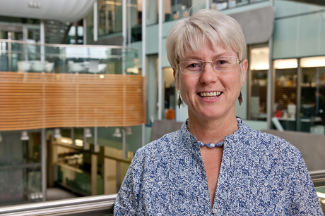
Professor Leann Tilley and her colleagues, Nick Klonis and Eric Hanssen, at the Bio21 Institute in Melbourne seem to spend a lot of their time designing and playing with the latest and greatest microscopy ‘super-toys’ to produce a myriad of weird and wonderful images of very, very small living things. While obviously a lot of fun for this multidisciplinary group that includes cell biologists, mathematicians, computer dudes and even physicists, the goal of this work is deadly serious.
For Tilley, it is part of her long-held research focus to better understand and control the ancient and ever-challenging problem of malaria, the parasitic infection that still kills up to 1 million people across the tropics each year, most of them children. And if recent results are anything to go by, the ‘fun’ is certainly time and money well spent.
The power and information provided by the latest techniques in super-resolution optical microscopy and electron tomography are finally revealing some of the malaria parasite’s long-held secrets.
A formidable adversary and weakening arsenal
Tilley has always been fascinated by the malaria parasite’s ability to undergo a remarkable series of morphological transformations during its life cycle through human and mosquito hosts. And à la Feynman’s famous advice, she believes that looking in great detail at these changes will help decipher the mechanisms underlying the parasite’s most intractable and problematic traits. In particular, the malaria parasite has developed resistance to nearly all of the antimalarial drugs thrown at it over many years.
“There are various drugs for treating malaria, but the parasite has developed resistance to most of these, so all the older ones like chloroquine are effectively useless,” said Tilley. “The most recent drug, an endoperoxide agent called artemisinin, still works although worrying evidence shows that resistance to this agent is also developing.”
Basically, none of the antimalarial drugs that are currently used are 100% effective - indeed, combinations of agents are now used to optimise efficacy and slow the spread of resistance. “These combinations are now working less well,” said Tilley, “and we desperately need replacement drugs because we know that the development of resistance is inevitable and we need to save the good ones like artemisinin for as long as possible.”
A major stumbling block to improving the existing antimalarial drugs is that scientists don’t really know how they work in the first place. The biological mechanisms of action for all the major players on the whole remain a mystery - even for the earliest stalwarts like chloroquine, and especially for artemisinin.
Targeting parasitic digestion
What is known is that for the malarial parasite to grow and prosper during its important proliferative stage inside the human host red blood cell (RBC), it needs a good source of amino acids … and what better source than the handiest around in the host’s own haemoglobin. It also needs to create a bit of growing room. Indeed, malaria parasites will ‘eat’ up to 75% of the host hemoglobin during an infection, using a stomach-like organelle referred to as the digestive vacuole.
Work from the Tilley lab and others showed that endoperoxide antimalarials, including artemisinin, are activated by the haem released during the haemoglobin breakdown. This led to studies implicating the digestive vacuole as an important site of artemisinin activity, with the parasite’s haemoglobin digestive process the drug target. More specifically, it was postulated that artemisinin is activated by interacting with the haem products, forming free radical species that react with things in the immediate vicinity of the parasite to induce cellular damage and, ultimately, cell death.
To investigate this, Tilley’s team started to use whole-cell electron tomography to look at the mechanics of haemoglobin digestion. “In this work, we prepare serial sections of the malaria organisms for viewing under an electron microscope and then acquire electron tomograms for each section using a series of tilt angles. The whole thing is then put back together computationally to build up a 3D picture of the structures inside the parasite and ideally the whole parasite or cell, but at the level of resolution enabled by electron microscopy (EM).”
Although incredibly labour- and time-intensive, Tilley found that this relatively new approach was fantastic for looking at the malaria parasite in human host cells … “and we could clearly see the digestive vacuole”. So, with the idea of the haemoglobin digestion and artemisinin mode of action in mind, they decided to look at it in more detail.
Watching and waiting pays off
“We started by morphologically tracking the digestive vacuole through the different stages of parasite development within the RBC, which takes about 48 hours,” explained Tilley. “Firstly, we were able to pinpoint the genesis of this organelle, which does not start until about a third of the way through the parasite RBC life cycle - so, for this time the parasite does not digest any haemoglobin.”
This finding clearly had implications in terms of artemisinin action and a possible means of resistance if haem production is necessary for the drug to work.
“Thus, by just looking at the different immature stages by EM, we could predict that the early stages of the parasite developing inside the RBC would be much less sensitive to artemisinin.” And when Tilley’s student, Stanley Xie, went back and did that experiment, this was indeed true.
“Also, when we biochemically stopped mature parasite stages from digesting haemoglobin, they became resistant to the presence of artemisinin,” said Tilley.
So if the parasite does not digest haemoglobin, artemisinin is inactive, confirming a mechanism by which the parasite could become drug-resistant. Tilley explained that this could work for the parasite because artemisinin is quite a ‘poor’ drug really, in that it only lasts in the bloodstream for a couple of hours - “so all the parasite has to do is find a way to hold off maturing for that time and then it can go in its merry way digesting haemoglobin and growing”.
Tilley then called on some mathematical colleagues, including James McCaw and Julie Simpson of the Melbourne School of Population and Global Health, to do some modelling studies of what would need to happen for these immature parasites to become resistant - that is, what level of resistance would they have to show for the treatment to fail. This very nice study confirmed that these immature parasites are the likely cause of artemisinin resistance.
“We now also have data coming in from the field telling us that our modelling data indeed reflects the real situation - when artemisinin fails to treat the parasite infection in patients, it fails because the immature parasites have become even more resistant than they already were. It seems that the ‘younger’ parasites can survive the presence of artemisinin for the few hours that the drug remains stable in the patient’s circulation.”
Straight to the source
Tilley is wasting no time translating their ‘high-end’ science into the real-life situation through collaboration with field colleagues. By collecting parasites from the field and adapting them to the laboratory for examination, the researchers can put their resistance hypothesis to the test. “We want to see how these parasites behave in our lab assays and down the microscopes to see if there is any difference in their development in terms of haemoglobin digestion.”
For these next steps, it is important to use parasites from areas where drug resistance is known to be a problem, as Tilley explained. “We get our samples from an area in Cambodia near the border with Thailand where there seems to be a kind of cradle of antimalarial drug resistance. In fact, it is the place where resistance first developed to chloroquine and other older antimalarial staples such as mefloquine, and now to artemisinin. The conditions there are obviously just right for the development of malarial resistance, although it is not entirely clear what those conditions are.”
It might be the nature of the area itself, something about the people that inhabit and pass through the area, something about the endemic parasite strains or, indeed, a combination of factors. “Whatever the reason, there is something about the parasite strains circulating in that area of Cambodia that makes it easier for them to develop resistance … and of course, from there the drug resistance mechanisms spreads all around the world as happened with chloroquine.”
Immediate benefits for treatment
According to Tilley, their electron tomography findings on the parasite life cycle and drug resistance could have immediate clinical applications for patients. One is in the area of drug dosing. Currently, artemisinin is administered as one dose per day for three consecutive days, a regimen largely designed on practical grounds to have the maximum effect.
“However, given what we now have realised about the mechanism of drug action and the timing of the parasite metabolism, this dosing schedule may not be optimal and perhaps only one of those doses will be present at the right time to have an effect while the other two might coincide with when the parasite is at its most resistant. Indeed, people are already looking at how we might be able to get better efficacy with existing drugs by changing the way they are administered and how such a change might be implemented in the field.”
Another implication is for current drug development efforts to synthesise longer-lived forms of endoperoxide antimalarials like artemisinin. “Our work would predict that such drugs will be much more effective to use against the early and resistant stages of the parasites - they can’t hold out forever and eventually will need to start digesting haemoglobin, thus enabling the drug to become active.”
For Tilley it is very exciting to see such high-end techniques like EM tomography used in a basic science context leading to a discovery that is potentially of immediate use for treating malaria in some of the world’s poorest populations.
Convergence working well
Tilley also sees the cross-disciplinary nature of her work, involving many Bio21 Institute scientists and partners, as one of the best things about it.
“We work with physicists to develop the imaging methods, mathematicians to do the modelling and computer scientists to improve the image analysis. It is true ‘convergence’ - this concept of bringing the biological and physical scientists together to work on big and important questions with a cross-disciplinary approach to generate novel and useful developments. Bio21 Institute really is a great place to do this and it seems to be working very well so far.”
**********************************************************************************
Views from Melbourne taking off
Tilley is currently Director of the ARC Centre of Excellence for Coherent X-ray Science (CXS), having recently taken the baton from Keith Nugent, who is now DVCR at La Trobe University. The centre brings physicists, chemists and biologists together to develop fundamentally new approaches to probing biological structures and processes. Tilley was instrumental (no pun intended) in developing the CXS approach and her laboratory has worked for many years on developing and implementing a number of new imaging modalities including coherent X-ray diffraction imaging, 3D electron tomography, cryo X-ray tomography and structured illumination microscopy. Tilley currently holds an ARC Australian Professorial Fellowship.
“At the Bio21 Institute, we now also have one of the first instruments in the world for live cell 3D Structure Illumination Microscopy (3D-SIM), and Trevor Smith is further developing the technology. This 3D-SIM BLAZE module from Applied Precision gives us an 8-fold increase in volume resolution compared with conventional light microscopes - that means you can see amazing details that we were not previously able to see in fluorescently labelled samples and collects the image data very rapidly to enable the 3D imaging of live cells at super resolution.”
**********************************************************************************
**********************************************************************************
The diverse application of EM tomography
EM tomography is basically a union of transmission electron microscopy, the latest advances in tomographic imaging and some high-end algorithmic and computing power. The result is some very impressive and useful images packed with novel and previously inaccessible information.
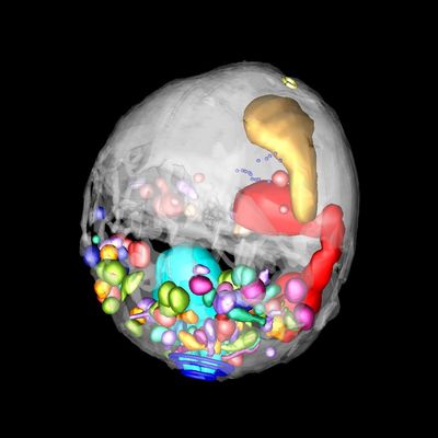
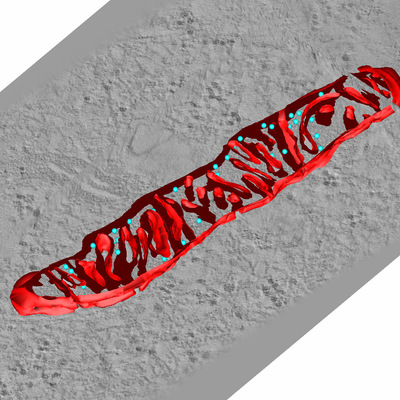
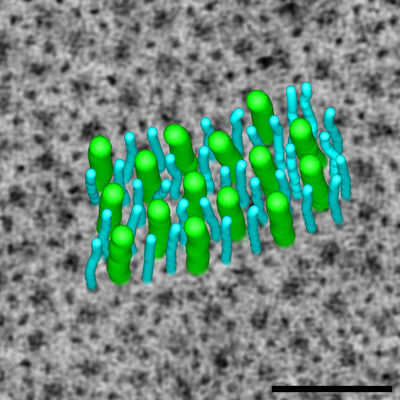
Other examples of tomographic imaging applications involving Eric Hanssen’s team at Bio21 include the structure of the surface layers of bacteria in dental health, the organisation and structure of nanoparticles and nanomaterials, the invasion of host red blood cells by malaria parasites (see image), the structure of milk component for the dairy industry, the effect of extracellular calcium phosphate crystals on inflammation, mitochondrial morphology in mitochondrial diseases (see image), the ultrastructure of collagen and muscle in muscular dystrophies and the organisation of the cell wall in plants, amongst others.


**********************************************************************************

AI-designed DNA switches flip genes on and off
The work creates the opportunity to turn the expression of a gene up or down in just one tissue...
Drug delays tumour growth in models of children's liver cancer
A new drug has been shown to delay the growth of tumours and improve survival in hepatoblastoma,...
Ancient DNA rewrites the stories of those preserved at Pompeii
Researchers have used ancient DNA to challenge long-held assumptions about the inhabitants of...


